

Etiological Pattern, Clinical Presentation, and Management Challenges of Proptosis in a Tertiary Hospital in South West Nigeria
Article Sidebar
Main Article Content
Abstract
Background: The etiology of proptosis is diverse ranging from orbital problem to infiltrative disease and spread from contiguous sites including nasopharynx, paranasal sinuses, and sometimes distant structures. It can also be part of systemic illness affecting multiple tissues and organs.
Aim: This study aims to determine the demographic pattern and etiology of proptosis in a tertiary health facility in South Western Nigeria and to discuss the management challenges.
Methods: This is a clinic‑based retrospective descriptive analysis of all patients that presented with proptosis at the Eye Clinic of Olabisi Onabanjo University Teaching Hospital Sagamu, Ogun state, Nigeria, over a 13‑year period from 2000 to 2012. The hospital records of patients was used which was analyzed using Statistical package for Social sciences version 15.
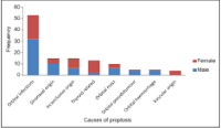
Results: A total of 175 cases of proptosis out of 15,266 new cases gave a hospital prevalence of 1.2%. The average age of the 138 patients analyzed was 37.8 years with a male to female ratio of 1:1. Children constituted 27.5%. Eighty‑one (58.7%) patients presented within 1 month of onset of proptosis. Twenty‑three (16.7%) had bilateral proptosis. Half of the studied population was secondary to orbital inflammation. The common causes of proptosis were infective 38.4%, mass/tumor 18.8%, noninfective inflammation 13%, and sinonasal diseases 10.9%. Eight (5.8%) were mucocele of paranasal sinuses. Thyroid‑related eye disease and proptosis of vascular etiology were common in females. Computerized tomographic scan of the orbit and/or sinus/cranium was done in 11.4% of the patients. Thirty‑seven (26.8%) patients defaulted.
Conclusion: Infective process is the most common cause of proptosis from orbital cellulitis. Majority were unilateral with no sex predilection. Proptosis due to thyroid eye disease and vascular abnormality were found mostly in females. The management challenges were poor record keeping and inadequate personnel. Despite the threat to life and vision posed by some etiology of proptosis, a large number of the patients were unable to fund investigation and treatment while others defaulted from the facility.
Downloads
Article Details
Section

This work is licensed under a Creative Commons Attribution-NonCommercial-ShareAlike 4.0 International License.
How to Cite


References
Kamminga N, Jansonius NM, Pott JW, Links TP. Unilateral proptosis: The role of medical history. Br J Ophthalmol 2003;87:370‑1.
Ahmed ME, Ahmed MK. Study of causes of proptosis in relation to demographic features among patients of El‑Minya University Hospital. El Minia Med Bull 2005;16:209‑15.
OgbeideE, TheophilusAO. Computed tomographic evaluation of proptosis in a Southern Nigerian tertiary hospital. Sahel Med J 2015;18:66‑70.
Siripurapu S, Raju TJ, Sekhar KC. A study on clinical correlation of orbital diseases interventional and non interventional diagnostic procedures. Indian J Public Health Res Dev 2016;7:164‑9.
Chaudhry IA, Shamsi FA, Elzaridi E, Al‑Rashed W, Al‑Amri A, Al‑Anezi F, et al. Outcome of treated orbital cellulitis in a tertiary eye care center in the middle east. Ophthalmology 2007;114:345‑54.
Majekodunmi S. Unilateral proptosis in Nigerians: Causes and differential diagnosis. Can J Ophthalmol 1982;17:203‑6.
Masud MZ, Babar TF, Iqbal A, Khan MT, Zaffar ul Islam, Khan MD. Proptosis: Etiology and demographic patterns. J Coll Physicians Surg Pak 2006;16:38‑41.
Satpute KK, Chingsuingamba Y. A retrospective analysis of presentation and management outcome of proptosis in a tertiary care centre of North‑East India – A case series. IOSR J Dent Med Serv 2013;5:30‑2.
Keche P, Naik SV, Nitnaware AZ, Mair M, Sakhare P, Satput S. A study of tumours giving rise to unilateral proptosis. Indian J Otolaryngol Head Neck Surg 2013;65:6‑13.
Lim NC, Sundar G, Amrith S, Lee KO. Thyroid eye disease: A Southeast Asian experience. Br J Ophthalmol 2015;99:512‑8.
Otulana TO, Sogebi OA, Onabolu OO, Ajibode HA, Bodunde OT. Proptosis as a presentation of nasopharyngeal malignancy in children. Niger Med J 2011;52:133‑7.
Sharma P, Tiwari PK, Ghimire PG, Ghimire P. Role of computed tomography in evaluation of proptosis. Nepal J Med Sci 2013;2:34‑7.
Gordon LK. Orbital inflammatory disease: A diagnostic and therapeutic challenge. Eye (Lond) 2006;20:1196‑206.
Khan NH, Moin M, Khain MA. Unilateral proptosis: A local experience. Biomedica 2004;20:114‑6.
DassRI, Maniar H, GurnaniD, Gohel DJ, Dave J, Desai N, et al. Proptosis presenting as a conmplication of sinusitis. Natl J Otorhinolaryngol Head Neck Surg 2013;1:13‑4.
Sindhu K, Downie J, Ghabrial R, Martin F. Aetiology of childhood proptosis. J Paediatr Child Health 1998;34:374‑6.
Sabharwal KK, Chouhan AL, Jain S. CT evaluation of proptosis. Indian J Rad Img 2006;16:683‑8.
Souza L, Caminha C. Orbital pseudotumor: A differential diagnosis of Graves’ ophthalmopathy. Arq Bras Endocrinol Metab 2011;55:85‑8.
Strianese D, Piscopo R, Elefante A, Napoli M, Comune C, Baronissi I, et al. Unilateral proptosis in thyroid eye disease with subsequent contralateral involvement: Retrospective follow‑up study. BMC Ophthalmol 2013;13:21.
Venugopal M, Sagesh M. Proptosis: The ENT surgeon’s perspective. Indian J Otolaryngol Head Neck Surg 2013;65 Suppl 2:247‑50.
Komolafe OO, Adeosun AA, Baiyeroju AM. Pattern of ophthalmic consult from the ear, nose and throat ward of a tertiary hospital. Niger J Ophthalmol 2009;17:11‑4.
Ghosh D, Khanna S, Baruah DK. Ophthalmological manifestations of ENT diseases: An overview. Indian J Otolaryngol Head Neck Surg 2013;65:197‑202.
Sikarwar V, Bisht RS, Kumar D, Shukla RK. Ophthalmic manifeastation in ENT disease surgical procedures. IOSR J Dent Med Sci 2013;11:87‑92.
Onakpoya OH, AdeoyeAO, Akinpelu OV. Cost‑related antibiotic dosage omissions‑challenge for orbital cellulitis management in resource poor communities. Orbit 2009;28:147‑52.
Eldesouky MA, Elbakary MA. Clinical and imaging characteristics of orbital metastatic lesions among Egyptian patients. Clin Ophthalmol 2015;9:1683‑7.